+1 713 980 8888
info@oilfindinternational.com
IMAGING SERVICES
Non-destructive imaging techniques provides a high-value complement for visualization and core assessment. Computed Tomography Scanners (CT), similar to medical applications, are used to capture a 3-Dimensional image of rocks. These images are taken when the cores arrive from the well sites and are still in their core barrels.
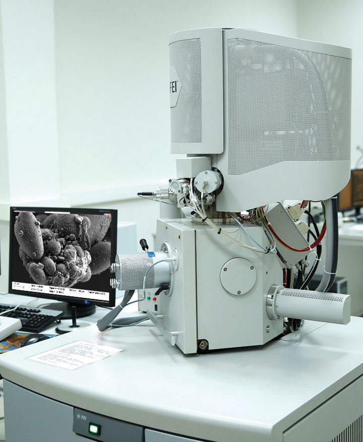
SEM
The Scanning Electron Microscope (SEM) images allow us to make a detailed characterization of the material surfaces with a focused beam of electrons. Different imaging techniques give qualitative and quantitative descriptions of:
- Representative features, such as: Overview images, texture, heterogeneity.
- High-Resolution features: Inter particle features, clay structures, pores, organics, diagenetic features are some of the details that are described using these high-resolution techniques. These permits to get insight on the phenomena occurring at the small scales.
- Mineralogy: An automatic classification of the different minerals provides detailed information on the composition of the different pore-scale structures.
- Processes assessment: Wettability changes (asphaltene precipitation), Formation Damage, detail understanding of processes through before/after imaging.
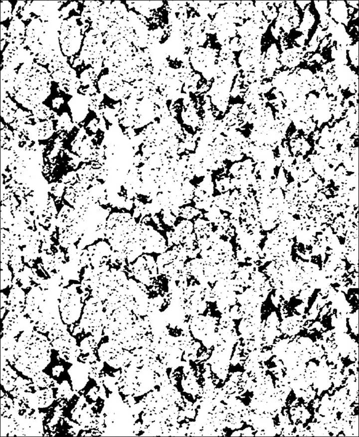
Micro-CT
Micro tomography is used to characterize the 3-Dimensional structures at the micrometer scales (20-200 times higher resolution than normal CT). This technology allows our clients to make a detailed characterization of plugs, which together with our Digital Rock tools allow to calculate properties such as: porosity, permeability, formation factor, capillary pressure, relative permeability’s, resistivity index, etc. This new technology allows us to quantify the effect of the pore-scale structure in global properties (e.g micro fractures, small vugs, clay contents, anomalies). MicroCT imaging can be applied to cores, plugs, unconsolidated samples, drill cuttings, etc. Some of the common services we perform for our clients are:
- Screening: High resolution imaging (10-20 um) for sample selection. With these images it is possible to distinguish the state of the material, fractures, select right areas for more detailed analysis, recognize feature, identify rock types, characterize heterogeneity, micro-scale features, contamination (drilling fluid invasion or other fluids), etc.
- Direct Image: Characterizing 3D features of pore-structures directly. This images allow us to make direct measures of porosity (when the pores are resolved) and perform different kind of simulations.
- Porosity Map: A set of different image techniques allows us to visualize the porosity distribution at the pore-scale with a resolution of a couple of micros.
- TOC (Total Organic Carbon): a set of different image techniques allows us to visualize the Organic content distribution.
- 4D imaging: Imaging before/after fluid invasion to analyze fluid distribution.
Conventional CT and Dual Energy CT (DECT)
Dual Energy tomography of cores provides a detailed 3-dimensional showing density and atomic number variations. Special clustering techniques allow us to convert this information into different lithotypes. Dual-energy scanning can also be used in fluid flow visualization studies. These interesting qualitative and quantitative results can improve the understanding of complex rocks, especially carbonates. Applications of DECT include subsample selection, direct high-resolution density and atomic number images, porosity, lithotypes, etc. The use of this new technology constitutes a vital part of the core analysis programs.
Digital Library
Once these 3-dimensional images are captured, they are archived in a digital library which can be accessed whenever the clients want. We build, maintain and manage digital repositories taking care of data Integrity, access, backups, format obsolescence, etc. The main advantage of this technology is to provide our clients with their rock images in the state as they were extracted, without the effect of the time as in conventional rock storage. You can access your rocks within years after they were extracted.
DIGITAL ROCK SERVICES
From direct measurements to complex simulations, the 2D and 3D images are used to quantify global properties of the rock material in different states (cores, plugs, cuttings, etc.). The images at different scales are combined to obtain accurate results, such as:
Petrophysical Properties
- Porosity
- Permeability
- Formation Factor
- Mechanical properties
Multi-Phase Properties
- Capillary Pressure
- Relative Permeability
- Resistivity Index
Complex Studies
- Heterogeneity characterization
- Sensitivity to Swi (Trapping curves)
- Sensitivity to wettability (contact angles)
- Characterization of EOR processes
- Unconventionals Rocks
- Tailored Digital Analysis
Our Address
Houston, Texas, USA
Call Us
+1 713 980 8888
Fax Us
+1 832 571 9908
Mail Us
info@oilfindinternational.com